Computed Tomography
Computed tomography (CT) is a fast and accurate imaging modality often used in emergency settings and trauma imaging. CT scans, with or without (or both) iodinated contrast are frequently used to image the brain, chest, abdomen and pelvis, but also have post-imaging reconstructive capabilities for detailed orthopedic imaging. It is now a standard imaging modality in emergency rooms to quickly assess patients. CT uses a series of X-ray images shot as the gantry rotates around the patient. Computer technology assembles these into into a dataset volume than can be slices on any access, or advanced visualization software can extract specific parts of the anatomy for study. Find more content specific to cardiac CT.

![Advanced artificial intelligence (AI) models can evaluate cardiovascular risk in routine chest CT scans without contrast, according to new research published in Nature Communications.[1] In fact, the authors noted, the AI approach may be more effective at identifying issues than relying on guidance from radiologists. Representative non-contrast CT slices for two patients (left), with super-imposed segmentations (right). One artificial intelligence (AI) model was used to segment a cardiac mask.](/sites/default/files/styles/top_stories/public/2024-04/screen_shot_2024-04-23_at_10.44.32_am.png.webp?itok=O8wgPFEZ)


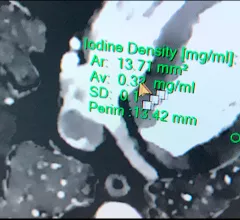
![Using computed tomography (CT) to perform coronary artery calcium (CAC) scoring can help identify symptomatic chest pain patients who do not require further testing, according to a new analysis published in Radiology.[1]](/sites/default/files/styles/top_stories/public/2024-03/screen_shot_2024-03-06_at_2.42.41_pm.png.webp?itok=jn2JGD8x)
 and professor of radiology at Mayo Clinic, shares his key takeaways from the SCCT 2022 conference. #SCCT #SCCT2022 #yesCCT")