Computed Tomography

Computed tomography (CT) is a fast and accurate imaging modality often used in emergency settings and trauma imaging. CT scans, with or without (or both) iodinated contrast are frequently used to image the brain, chest, abdomen and pelvis, but also have post-imaging reconstructive capabilities for detailed orthopedic imaging. It is now a standard imaging modality in emergency rooms to quickly assess patients. CT uses a series of X-ray images shot as the gantry rotates around the patient. Computer technology assembles these into into a dataset volume than can be slices on any access, or advanced visualization software can extract specific parts of the anatomy for study. Find more content specific to cardiac CT.
SCCT President Ed Nicol, MD, explains key trends trends in cardiac CT imaging at the 2023 meeting. #SCCT #SCCT23 #SCCT2023

Key trends and takeaways in cardiac CT at SCCT 2023

Several key trends at the 2023 annual meeting of the Society of Cardiovascular Computed Tomography were outlined by society President Ed Nicol, MD, including AI, reimbursement issue, business management discussions, and a growing interest in cardiac CT leading to record attendance this year.

August 1, 2023
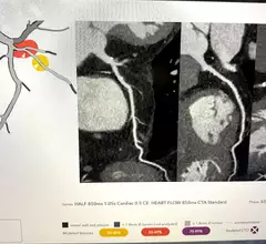
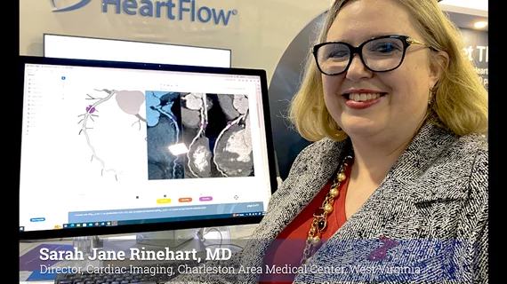
HeartFlow's RoadMap technology where AI reads the image in a first pass to highlight areas of interest with for the radiologist or cardiologist on coronary CT scan. The technology was shown to reduce reading time by 25%.

Three studies show value of AI to speed cardiac CT reads and soft plaque evaluation 

AI to help interpret cardiac CT was shown to reduce exam read times to improve workflow efficiency, and improve patient diagnostics with additional information on soft plaques. Data was presented in three HeartFlow AI technology studies at SCCT 2023.

July 31, 2023
scad_1.jpg

Photon-counting technology offers new opportunities in imaging high-risk CAD patients

Coronary CT angiography is commonly used to assess patients at low or intermediate risk of CAD but is less effective in high-risk patients with an increased presence of coronary calcifications and stents due to the “blooming” effect they have on imaging.

June 21, 2023
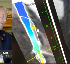
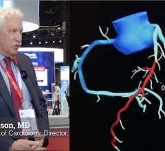
Ed Nicol, MD, consultant cardiologist and honorary senior clinical lecturer with Kings College London and president-elect of the Society of Cardiovascular Computed Tomography (SCCT), explained artificial intelligence (AI) in cardiac CT is here to stay and its use is expanding. He noted that one AI-based algorithm is already included in recent cardiology guidelines and more will likely follow. #SCCT

Cardiac imagers need to understand AI as it enters clinical use and ACC guidelines

Most FDA-cleared AI algorithms are related to radiology and cardiology, meaning radiologists and cardiologists need to make an effort to learn how these technologies work.

February 15, 2023
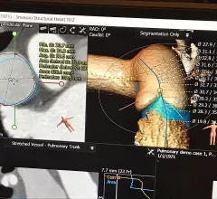
An example of virtual pulmonary valve implant planning software showing a virtual transcatheter pulmonary valve with measurements implnated in the virtual anatomy of a congenital heart patient. This was part of a training class at the Society of Cardiovascular Computed Tomography (SCCT) 2022 meeting.

FDA makes clearance easier for cardiac device implant simulation software

The FDA is reclassifying interventional cardiovascular implant simulation software into the class II (special controls) regulatory category, which will make it easier for the technology to pass regulatory approval. 

January 4, 2023
James Udelson, MD, from Tufts and an investiogator in the PRECISE trial, explains how FFR-CT combined with coronary CT outperformed the standard of care in patients with stable chest pain, or low-risk patients with suspected coronary involvement.

VIDEO: PRECISE trial shows cardiac CT with FFR-CT significantly improves patient outcomes

James Udelson, MD, an investigator involved with the PRECISE trial, explained how patient outcomes were improved by 70% from the current standard of care.

November 8, 2022
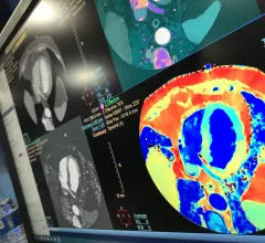
A spectral CT image of the heart showing ischemia in the myocardium by mapping the iodine density in the tissues as a surrogate for bloodflow. Example is from Philips Healthcare, displayed at the 2022 ACC meeting. Example of cardiac perfusion imaging. #SCCT

VIDEO: The many benefits of spectral CT for cardiovascular imaging

Suhny Abbara, MD, editor of Radiology: Cardiothoracic Imaging and chief of cardiothoracic imaging for University of Texas Southwestern Medical Center, discusses how spectral computed tomography (CT) can help both cardiac and general CT imaging.

November 8, 2022
medical billing_5.jpg

Neuroimaging recedes behind other cost compilers in stroke care

Contrary to older research that showed neuroimaging emerging as the single most dominating cost contributor in ischemic stroke care for older Americans, a new study shows treatment and other line items account for bigger slices of the bill.

November 8, 2022

Around the web

Eleven medical societies have signed on to a consensus statement aimed at standardizing imaging for suspected cardiovascular infections.

Kate Hanneman, MD, explains why many vendors and hospitals want to lower radiology's impact on the environment. "Taking steps to reduce the carbon footprint in healthcare isn’t just an opportunity," she said. "It’s also a responsibility."

Trimed Popup
Trimed Popup