Computed Tomography

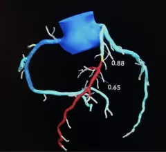
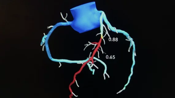
Cardiac computed tomography (CT) has become a primary cardiovascular imaging modality in the past 20 years, and was recommended as a 1A recommendation in the 2021 chest pain assessment guidelines. CT calcium scoring has became a primary risk assessment for coronary artery disease and whether patients should be on statins. Coronary CT angiography (CCTA) is used to for anatomical assessment of the arteries for plaque burden and to identify areas of blockage that may cause ischemia and heart attacks. Additional use of contrast CT perfusion or fractional flow reserve CT (FFR-CT) can offer physiological information on the function of the heart. CT plays a primary role in structural heart assessments for heart valves, repair of congenital defects and left atrial appendage occlusion (LAAO) for both pre-procedure planning and procedural guidance. Find more news on general radiology CT use.
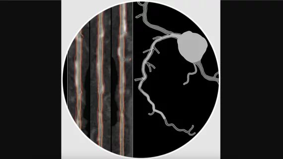
HeartFlow Plaque Analysis. A European Heart Journal – Cardiovascular Imaging study compared the noninvasive Plaque Analysis technology developed by HeartFlow, which uses advanced AI algorithms to evaluate coronary CT angiography (CCTA) images, with coronary plaque assessments achieved by using intravascular ultrasound (IVUS). The REVEALPLAQUE trial included data from 237 patients. Plaque Analysis showed strong correlation and high accuracy when compared with IVUS.

AI-powered coronary plaque assessments show ‘strong agreement’ with IVUS

HeartFlow's noninvasive Plaque Analysis technology, which uses AI to evaluate CCTA images, delivered assessments that mostly lined up with IVUS results. 

May 16, 2024
Performing CT-derived fractional flow reserve (CT-FFR) before transcatheter aortic valve replacement (TAVR) improves the accuracy of coronary CT angiography (CCTA) and helps limit unneeded invasive coronary angiography (ICA), according to a new study published in JACC: Cardiovascular Interventions. Heartflow

AI-based CAD assessments dramatically improve vascular surgery outcomes

PAD patients evaluated with HeartFlow's noninvasive FFRCT Analysis technology prior to surgery experienced much better outcomes, including a 63% lower risk of all-cause mortality after five years. 

May 14, 2024
Remote ECG access on a smart phone via the Viz.ai Cardio Suite, which enables access to dynamic ECG, echo, MRI, CT images and reports. It also enables automated detection on imaging to alert care teams for STEMI, hypertrophic cardiomyopathy, pulmonary embolism, aortic dissection and abdominal aortic aneurysm, and heart failure.

Cardiology ranked No. 2 among all specialties with 122 FDA-cleared AI models

Only radiology is associated with more FDA-cleared AI algorithms than cardiology, according to new federal data. 

May 14, 2024
Interview with Nehal Mehta, MD, University of Pennsylvania, who explains how coronary inflammation can be seen using AI on cardiac CT scans to better risk stratify patients and begin preventive drug therapy.

AI helps cardiologists track new drug's effect on inflammation

The combination of AI and CT helped Nehal Mehta, MD, and colleagues track the performance of a new drug designed to target coronary inflammation. 

May 7, 2024
Sarah Jane Rinehart, MD, director of cardiac imaging, Charleston Area Medical Center, Charleston West Virginia, as been using the FDA-cleared RoadMap artificial intelligence algorithm from HeartFlow in studies and in clinical used since it was cleared and said it helps cardiologists in several ways. #ACC #ACC24 #ACC2024 #Heartflow #AIhealth

AI improves CT assessments, boosts care for real-world heart patients

Automated AI-generated measurements combined with annotated CT images can improve treatment planning and help referring physicians and patients better understand their disease, explained Sarah Jane Rinehart, MD, director of cardiac imaging with Charleston Area Medical Center.

April 26, 2024
Advanced artificial intelligence (AI) models can evaluate cardiovascular risk in routine chest CT scans without contrast, according to new research published in Nature Communications.[1] In fact, the authors noted, the AI approach may be more effective at identifying issues than relying on guidance from radiologists. Representative non-contrast CT slices for two patients (left), with super-imposed segmentations (right). One artificial intelligence (AI) model was used to segment a cardiac mask.

AI predicts cardiovascular risk during CT scans—no invasive tests or contrast required

Two advanced algorithms—one for CAC scores and another for segmenting cardiac chamber volumes—outperformed radiologists when assessing low-dose chest CT scans. 

April 23, 2024
Performing CT-derived fractional flow reserve (CT-FFR) before transcatheter aortic valve replacement (TAVR) improves the accuracy of coronary CT angiography (CCTA) and helps limit unneeded invasive coronary angiography (ICA), according to a new study published in JACC: Cardiovascular Interventions. Heartflow

New cardiac imaging strategy could reduce ICA, PCI rates

Radiologists with Massachusetts General Hospital found that the selective use of cardiac CT and AI-based CAD evaluations could make a significant impact on patient care. 

April 16, 2024
The V-Wave intra-atrial shunt to treat heart failure on display on the expo floor at ACC.24. The device was used in one of the late breaking trials at ACC. DF 3

PHOTO GALLERY: Highlights from ACC.24 in Atlanta

ACC.24, the American College of Cardiology's annual meeting in Atlanta, featured the latest in cardiovascular research and technologies. Representatives from Cardiovascular Business were there in person to take in the excitement. 

April 16, 2024

Around the web

Eleven medical societies have signed on to a consensus statement aimed at standardizing imaging for suspected cardiovascular infections.

Kate Hanneman, MD, explains why many vendors and hospitals want to lower radiology's impact on the environment. "Taking steps to reduce the carbon footprint in healthcare isn’t just an opportunity," she said. "It’s also a responsibility."

Trimed Popup
Trimed Popup