Nuclear Cardiology

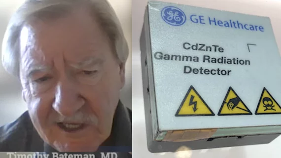
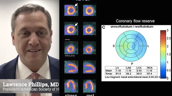
Single photon computed tomography (SPECT) and positron emission tomography (PET) molecular imaging are used as primary cardiac imaging modalities to evaluate the function of the heart. It uses radioactive isotopes attached to sugars that are metabolized by cardiomyocytes. This creates an image of the metabolic activity of the heart and shows areas of ischemia or infarct. Other radiotracers can image the heart to diagnosis cardiac amyloidosis and sarcoidosis. 

PET technique visualizes amyloid deposits in heart

PET with 11C-PIB provides a noninvasive method for visualizing amyloid deposits in the heart, according to a study published in the February issue of the Journal of Nuclear Medicine. The researchers suggest that 11C-PIB eventually may be used in the clinical setting as both a diagnostic tool and a treatment follow-up method.

February 10, 2013

ASNC Releases Dose-Reduction Guidelines for Nuclear Cardiology

Technological improvements in image acquisition and software processing in nuclear cardiology should allow physicians to shave patient imaging times dramatically or cut radiation doses fourfold, according to a new preferred practice statement from the American Society of Nuclear Cardiology (ASNC).

March 2, 2012

Around the web

Eleven medical societies have signed on to a consensus statement aimed at standardizing imaging for suspected cardiovascular infections.

Kate Hanneman, MD, explains why many vendors and hospitals want to lower radiology's impact on the environment. "Taking steps to reduce the carbon footprint in healthcare isn’t just an opportunity," she said. "It’s also a responsibility."

Trimed Popup
Trimed Popup