Harvard engineers have used a new manufacturing method, focused rotary jet spinning (FRJS), to recreate the helical structure of human heart muscles. The breakthrough could prove to be a significant moment in the history of heart transplant technology, putting specialists one step closer to fully constructing a human heart that can then be transplanted into a living patient.
The team at Harvard shared its findings in the journal Science, noting that FRJS “offers a streamlined approach to fabricating tissues and organs” while providing a fresh perspective on how they function.[1]
“This work is a major step forward for organ biofabrication and brings us closer to our ultimate goal of building a human heart for transplant,” senior author Kit Parker, PhD, a professor of bioengineering and applied physics at the Harvard John A. Paulson School of Engineering and Applied Sciences (SEAS), said in a statement.
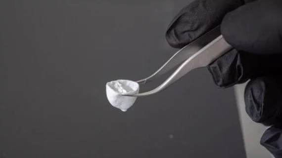
Parker et al. used FRJS—which is faster and much more efficient than 3D printing—to develop what they believe to be the first biohybrid model of human ventricles with helically aligned beating cardiac cells. The team hoped to test Edward Sallin’s 1969 theory that the heart’s helical design is closely associated with a patient’s ejection fractions.
"Since 2003, our group has worked to understand the structure-function relationships of the heart and how disease pathologically compromises these relationships,” Parker added. “In this case, we went back to address a never tested observation about the helical structure of the laminar architecture of the heart. Fortunately, professor Sallin published a theoretical prediction more than a half century ago and we were able to build a new manufacturing platform that enabled us to test his hypothesis and address this centuries-old question.”
After the FRJS produced ventricle models, rat cardiomyocyte or human stem cell-derived cardiomyocyte cells were added. Within roughly one week, the authors noted, thin layers of beating tissue covered the scaffold of the ventricles—and the cells closely followed the fibers’ alignment, going through the same “twisting or wringing” motion seen in actual human hearts.
The SEAS team plans to continue its work in biofabrication and has already started considering the commercial potential of this technology in other areas, including food packaging.
Related Heart/Organ Modeling Content:
3D-printed heart valve models have the feel and look of the real thing
Worth the wait: After years of trying, researchers 3D print a working heart pump with human cells
3D printing could help prevent paravalvular leak in TAVR patients
3D cardiac modeling solution for VT ablation receives FDA clearance
Reference:

Michael has more than 16 years of experience as a professional writer and editor. He has written at length about cardiology, radiology, artificial intelligence and other key healthcare topics.