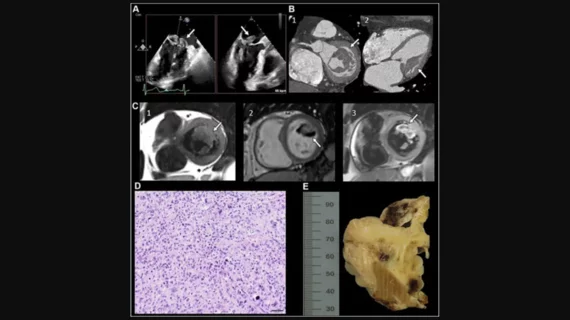
The winning images: A 75-year-old female patient with an incidental finding at transesophageal echocardiography of a lobulated intracardiac mass on the mitral valve (Fig 1A). Comprehensive characterization of the mass using multimodality imaging assessment was performed. With cardiac CT, the precise extent of the mass was detailed with involvement of both mitral valve leaflets and extension to the papillary muscles but without infiltration into the adjacent myocardium (Fig 1B). Multiparametric contrast-enhanced cardiac MRI further characterized the complexity of the mass, showing minimal perfusion and late gadolinium enhancement, with features suggestive of multiple adherent thrombi (Fig 1C). Biopsy and histopathologic assessment confirmed the mass to be a high-grade undifferentiated pleomorphic sarcoma (Fig 1D). Image/caption courtesy of RSNA.