Representative non-contrast CT slices for two patients (left), with super-imposed segmentations (right). One artificial intelligence (AI) model was used to segment a cardiac mask (magenta line) and coronary artery calcium (red). A second AI model segmented left ventricular myocardium (purple), left atrial (green), left ventricle (light red), right ventricle (blue) and right atrial (yellow) volumes. Images/caption courtesy of Slomka et al. and Nature Communications.
Two advanced algorithms—one for CAC scores and another for segmenting cardiac chamber volumes—outperformed radiologists when assessing low-dose chest CT scans.
Researchers asked ChatGPT to make treatment decisions for patients with severe aortic stenosis, comparing its answers to the recommendations of a full heart team.
The three-day event will include sessions about sexual activity among heart patients, depression, energy drinks, stimulants, vaccines and more. The festivities begin Thursday, April 25, in Greece.
“The alleged conduct of this physician is so egregious, only the permanent revocation of his license could adequately protect the public from the risks posed by his return to practice," one official said.
AskBio, acquired by Bayer for more than $2 billion in 2020, is enrolling patients for a new clinical trial focused on the therapy's effectiveness in adult patients with non-ischemic cardiomyopathy and heart failure.
Alexander Fanaroff, MD, said the late-breaking BE ACTIVE clinical trial presented at ACC.24 offers a blueprint for how to get patients to be more physically active.
Stroke is typically seen as the biggest danger for patients after they receive an AFib diagnosis. This study, however, suggests heart failure could be an even bigger threat.
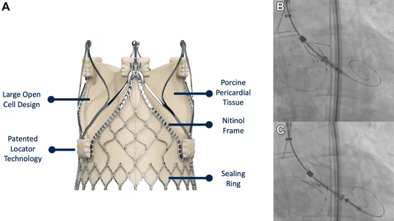
The JenaValve Trilogy system allows for placement in noncalcific pure aortic regurgitation (A). Using a transcaval access, the system was positioned above the aortic valve (B). It was then released, clipping itself to the valve using the dedicated locator mechanism (C). Images/caption courtesy of Curio et al., JACC: Case Reports.
The 65-year-old male patient presented with a long medical history and many comorbidities, making surgery too risky.
![Advanced artificial intelligence (AI) models can evaluate cardiovascular risk in routine chest CT scans without contrast, according to new research published in Nature Communications.[1] In fact, the authors noted, the AI approach may be more effective at identifying issues than relying on guidance from radiologists. Representative non-contrast CT slices for two patients (left), with super-imposed segmentations (right). One artificial intelligence (AI) model was used to segment a cardiac mask.](/sites/default/files/styles/top_stories/public/2024-04/screen_shot_2024-04-23_at_10.44.32_am.png.webp?itok=O8wgPFEZ)