Magnetic Resonance Imaging

Magnetic resonance imaging (MRI) is the gold standard imaging modality for soft tissues. It produces detail cross-sectional images of soft tissue and bone anatomy, including muscles, tendons, ligaments, brain and organs, without the use of ionizing radiation. In addition to orthopedic imaging, MRI is also used for heart, brain and breast. MRI uses gadolinium contrast in many exams to highlight tissues and blood vessels, which enhances images and offers better diagnostic quality. It can also be used in conjunction with PET scans. How does MRI work? MR creates images by using powerful magnets to polarize hydrogen atoms in water (the body is made of of more than 80% water) so they face in one direction. A radiofrequency pulse is then used to ping these atoms, causing them to wobble, or resonate. The MRI coils detect this and computers can assemble images from the signals. Basic MRI scans will focus on the resonance of fat and water in two different sequences, which highlight and contrast different features in the anatomy.
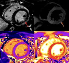
COVID-19 vaccine associated myocarditis on short-axis 1.5T MRI images of a 19-year-old man who presented with chest pain three days following the second dose of an mRNA COVID-19 vaccine.

New cardiac MRI analysis offers updated insight into long-term impact of vaccine-related myocarditis

Months after their initial myocarditis diagnosis, late gadolinium enhancement (LGE) was observed in 62% of patients on follow-up cardiac MRI.

May 4, 2022

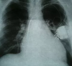
Cardiac implantable electronic devices can be safely reprogrammed to MRI-safe mode via remote operator

In 209 study participants, the remote reprogramming resulted in a successful, event-free MRI scan for every patient.

May 4, 2022
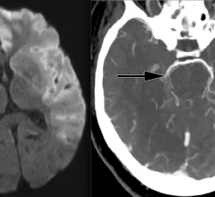
Ischemic stroke CT imaging. Images courtesy of RSNA

Stroke care still lags among certain Medicare populations

Despite increases of 250% for CTA and 428% for CTP, researchers observed lower rates of neuroimaging utilization among rural residents, older individuals (80 years and above), women and Black patients.

April 27, 2022
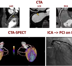
A figure from the 2022 CAD non-invasive imaging guidelines showing a comparison of computed tomography angiography (CTA) and a SPECT-CT vs. an invasive angiogram from the cath lab showing the same blockage in a coronary artery.

New multi-society recommendations highlight role of non-invasive imaging in evaluating coronary artery disease

A new, multi-society document, "Non-Invasive Imaging in Coronary Syndromes," focuses on how multiple imaging techniques can evaluate different aspects of coronary artery disease (CAD), all without the need for invasive angiograms.

April 21, 2022
Collaborators from a number of international centers, led by the European Molecular Biology Laboratory-European Bioinformatics Institute, published their findings Wednesday in Nature. Using MRI scans, along with morphology and genetic data, they showed that this network of heart muscle fibers—known as the trabeculae—can influence the heart’s performance.

Thousands of MRI scans help experts understand 500-year-old heart mystery first described by Leonardo da Vinci

Artificial intelligence also played a large role in discovering how this intricate network of muscle fibers can influence health, experts explained in Nature.

August 19, 2020
stroke

What we can learn about stroke from a simple MRI technique

Using MRI scans to measure iron content can help specialists learn more about stroke-related damage to the brain, according to a new study published by Radiology. Should such measurements be required after patients suffer a stroke?

March 14, 2019
""

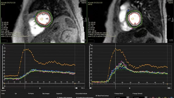
Cardiac MRI-derived T2 mapping may help heart failure patients

Researchers used T2 mapping taken from weekly cardiac MRIs to help identify cardiotoxicity at an early stage, according to results of a pig study published Feb. 18 in the Journal of the American College of Cardiology. The findings could help cancer patients at risk of chemo-induced heart failure.

February 22, 2019

T2 mapping may uncover cardiotoxic marker early enough to prevent heart failure

T2 mapping derived from weekly cardiac MRIs helped researchers identify cardiotoxicity at an early and reversible stage, a finding which may have implications for cancer patients at risk of chemotherapy-induced heart failure.

February 21, 2019

Around the web

Eleven medical societies have signed on to a consensus statement aimed at standardizing imaging for suspected cardiovascular infections.

Kate Hanneman, MD, explains why many vendors and hospitals want to lower radiology's impact on the environment. "Taking steps to reduce the carbon footprint in healthcare isn’t just an opportunity," she said. "It’s also a responsibility."

Trimed Popup
Trimed Popup