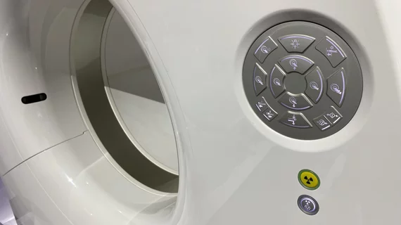
Magnetic Resonance Imaging

Magnetic resonance imaging (MRI) is the gold standard imaging modality for soft tissues. It produces detail cross-sectional images of soft tissue and bone anatomy, including muscles, tendons, ligaments, brain and organs, without the use of ionizing radiation. In addition to orthopedic imaging, MRI is also used for heart, brain and breast. MRI uses gadolinium contrast in many exams to highlight tissues and blood vessels, which enhances images and offers better diagnostic quality. It can also be used in conjunction with PET scans. How does MRI work? MR creates images by using powerful magnets to polarize hydrogen atoms in water (the body is made of of more than 80% water) so they face in one direction. A radiofrequency pulse is then used to ping these atoms, causing them to wobble, or resonate. The MRI coils detect this and computers can assemble images from the signals. Basic MRI scans will focus on the resonance of fat and water in two different sequences, which highlight and contrast different features in the anatomy.
For almost nine years since the adoption of the Protecting Access to Medicare Act (PAMA) established the requirement for appropriate use criteria (AUC) to receive payment for advanced diagnostic imaging, the American Society of Nuclear Cardiology (ASNC) has lobbying Congressional leaders change or repeal AUC mandate. Those efforts have paid off in the 2024 Medicare Physician Fee Schedule final rule, where AUC program has been paused and all of its regulations were rescinded. Photo by Dave Fornell.

Medicare drops AUC requirement for advanced imaging, ASNC celebrates

"This was an unneeded burden, which was solely adding to the administrative hassles of medicine," said American Society of Nuclear Cardiology President Larry Phillips.

April 8, 2024
Kate Hanneman, MD, University Of Toronto, explains why vendors and hospitals are increasingly discussing lowing their carbon footprint by starting with radiology. 

What does radiology have to do with climate change?

Kate Hanneman, MD, explains why many vendors and hospitals want to lower radiology's impact on the environment. "Taking steps to reduce the carbon footprint in healthcare isn’t just an opportunity," she said. "It’s also a responsibility."

March 1, 2024
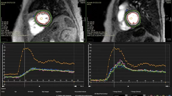
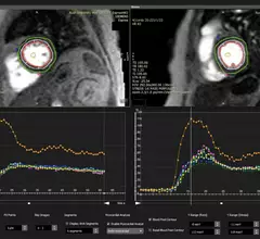
An example of cardiac MRI myocardial blood flow reserve (MBFR) imaging. Image courtesy of https://doi.org/10.3390/diagnostics10090679

Imaging group thrilled with new CPT codes for MBFR measurements

The Society for Cardiovascular Magnetic Resonance applauded two new CPT Category III codes for myocardial blood flow reserve (MBFR) assessments.

February 16, 2024

Multidisciplinary quality improvement initiative significantly improves on-time starts for cardiac MRI

Such delays can result in longer patient fasts, extended wait times, and poor synchronization of anesthesia induction, researchers detailed in JACR

October 16, 2023
gadolinium, GBCAs

Generic substitute for gadolinium-based contrast agent Gadavist hits the market

The contrast agent, simply called Gadobutrol Injection, has been approved by the U.S. Food and Drug Administration and is now available in the United States.

August 9, 2023
Medtronic just launched its MRI Care Pathway, a new system that can streamline the process of completing MRI scans for patients with Medtronic MRI compatible cardiac devices.

Medtronic streamlines MRI screening, scanning for patients with implanted cardiac devices

Medtronic launched its MRI Care Pathway, a new system that can streamline the process of completing MRI scans for patients with compatible cardiac devices.

April 4, 2023
medical billing_5.jpg

Neuroimaging recedes behind other cost compilers in stroke care

Contrary to older research that showed neuroimaging emerging as the single most dominating cost contributor in ischemic stroke care for older Americans, a new study shows treatment and other line items account for bigger slices of the bill.

November 8, 2022
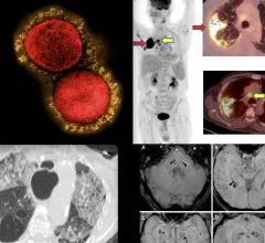
COVID-19 medical imaging examples of various clinical presentations. SARS-CoV-2 clinical imaging presentations.

PHOTO GALLERY: What does COVID-19 look like on medical imaging?

This image gallery shows what the various clinical presentations associated with the COVID-19 virus that have been documented during the coronavirus pandemic.

August 9, 2022

Around the web

Eleven medical societies have signed on to a consensus statement aimed at standardizing imaging for suspected cardiovascular infections.

Kate Hanneman, MD, explains why many vendors and hospitals want to lower radiology's impact on the environment. "Taking steps to reduce the carbon footprint in healthcare isn’t just an opportunity," she said. "It’s also a responsibility."

Trimed Popup
Trimed Popup