Magnetic Resonance Imaging

Cardiac magnetic resonance imaging MRI is used as both a functional and anatomical cardiac imaging test. It offers excellent soft tissue detail and the ability to quantify cardiac function. MRI scans can be performed with or without gadolinium contrast depending on what information is needed. Unlike computed tomography (CT), MRI does not use X-ray radiation, but patients with metal implants may have contraindications for MRI use because MR will heat up most metal objects. MRI exams usually take much longer than CT scans. How does MRI work? MR creates images by using powerful magnets to polarize hydrogen atoms in water (the body is made of of more than 80% water) so they face in one direction. A radiofrequency pulse is then used to ping these atoms, causing them to wobble, or resonate. The MRI coils detect this and computers can assemble images from the signals. Basic MRI scans will focus on the resonance of fat and water in two different sequences, which highlight and contrast different features in the anatomy.
New imaging protocols proposed to curb rise of cardiovascular infections

Eleven medical societies have signed on to a consensus statement aimed at standardizing imaging for suspected cardiovascular infections.

March 12, 2024
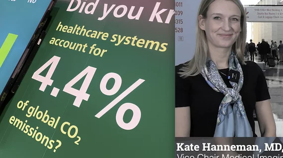
Kate Hanneman, MD, University Of Toronto, explains why vendors and hospitals are increasingly discussing lowing their carbon footprint by starting with radiology. 

What does radiology have to do with climate change?

Kate Hanneman, MD, explains why many vendors and hospitals want to lower radiology's impact on the environment. "Taking steps to reduce the carbon footprint in healthcare isn’t just an opportunity," she said. "It’s also a responsibility."

March 1, 2024
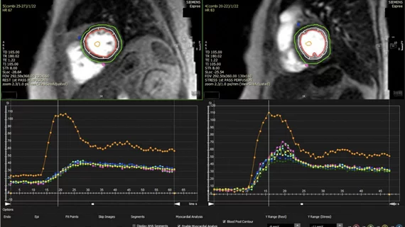
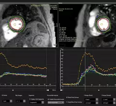
An example of cardiac MRI myocardial blood flow reserve (MBFR) imaging. Image courtesy of https://doi.org/10.3390/diagnostics10090679

Imaging group thrilled with new CPT codes for MBFR measurements

The Society for Cardiovascular Magnetic Resonance applauded two new CPT Category III codes for myocardial blood flow reserve (MBFR) assessments.

February 16, 2024
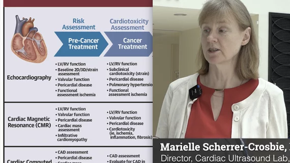
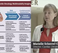
Video interview with Marielle Scherrer-Crosbie, MD, Hospital of the University of Pennsylvania, who explains details of creating a cardio-oncology program, whoi should be involved and the role of cardiac imaging.

Key things to remember when creating a cardio-oncology program

Marielle Scherrer-Crosbie, MD, explains what is needed to create a cardio-oncology program, and the role played by cardiac imagers.

February 12, 2024
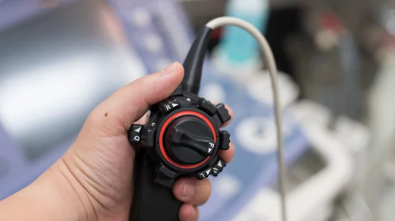
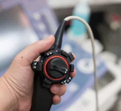
transesophageal echocardiography (TEE)

New expert guidelines: Start PHV evaluations with echocardiography, but other imaging modalities can provide value

New guidelines from the American Society of Echocardiography, made in collaboration with the Society for Cardiovascular Magnetic Resonance and Society of Cardiovascular Computed Tomography, represent an update of the group's original recommendations from 2009.

January 9, 2024
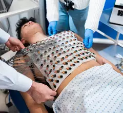
UCL Imaging vest screening cardiac conditions

New vest developed by cardiologists uses advanced heart imaging to screen for sudden cardiac arrest

“We identified a problem in cardiology,” one cardiologist explained. “Heart imaging has made remarkable progress in recent decades, but the electrics of the heart have eluded us."

December 19, 2023
João Cavalcante, MD, Minneapolis Heart Institute, spoke at the Radiological Society of North America (RSNA) 2023 meeting to try and get more radiologists interested in cardiac imaging to help fill the rising need for cardiac imagers on structural heart teams and a growing number of other types of heart and acute care teams.

Filling the crucial role of multimodality imagers on the heart team

João Cavalcante, MD, spoke at RSNA 2023 about key topics and tried to get more radiologists interested in cardiac imaging.

December 15, 2023
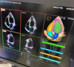
Example of a totally AI driven echocardiography workflow on the new Siemens Origin ultrasound system unveiled in 2023. The AI did all the work on this screen, taking a 3D echo exam and automatically segmenting the anatomy, contoured all the chambers, found the ideal views to display and then calculated all the measurements in seconds. Photo by Dave Fornell at TCT 2023. 

Cardiology now has more than 100 FDA cleared AI algorithms; experts say that is just the beginning

Cardiology makes up 10% of the 692 market-cleared clinical AI algorithms in the FDA’s latest update on the number of patient-facing AI now commercialized in the U.S. Experts share their thoughts on how it is being used.

December 14, 2023

Around the web

Eleven medical societies have signed on to a consensus statement aimed at standardizing imaging for suspected cardiovascular infections.

Kate Hanneman, MD, explains why many vendors and hospitals want to lower radiology's impact on the environment. "Taking steps to reduce the carbon footprint in healthcare isn’t just an opportunity," she said. "It’s also a responsibility."

Trimed Popup
Trimed Popup