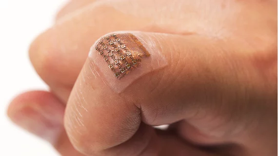
Scientists have developed a wearable ultrasound device to measure central blood pressure (BP). It performed as well as a current noninvasive technique upon testing, according to a press release.
The stretchable patch, which is only 240 micrometers thick, can continuously monitor central blood pressure in arteries up to four centimeters below the skin.
“Wearable devices have so far been limited to sensing signals either on the surface of the skin or right beneath it. But this is like seeing just the tip of the iceberg,” Sheng Xu, senior study author and a professor of nanoengineering at the University of California, San Diego, said in the release. “By integrating ultrasound technology into wearables, we can start to capture a whole lot of other signals, biological events and activities going on way below the surface in a non-invasive manner.”
Another coauthor said the device could be particularly useful in the operating room during complex cardiopulmonary procedures.
When most people think of blood pressure tests, they picture the arm cuff device, but that’s used to measure peripheral blood pressure. Central BP is the pressure in the central vessels that connect the heart director to other major organs in the body, according to the release.
The current standard for measuring central BP requires inserting a catheter into a patient’s blood vessel and guiding it to the heart, the release stated. A noninvasive method also exists, but in those cases a tonometer must be held steady, at a proper angle and with consistent pressure—making it likely readings could differ from test to test and from clinician to clinician.
“Even with the proper technique, if you move the tonometer tip just a millimeter off, the data get distorted,” said first author Chonghe Wang, also of UC San Diego.
The researchers tested the wearable patch against a tonometer and found their innovation was more consistent and precise, with its recordings also comparable to a traditional ultrasound probe. Ultrasound waves continuously record the diameter of blood vessels pulsing beneath the skin, providing information on a patient’s cardiovascular health and blood supply, according to the release.
However, they noted several improvements must be made before the device can be used in clinical practice, such as integrating a power source, data processing units and wireless communication.
“Right now, these capabilities have to be delivered by wires from external devices,” Xu said. “If we want to move this from benchtop to bedside, we need to put all these components on board.”
Xu and colleagues described their early experience with the ultrasound patch in Nature Biomedical Engineering.

