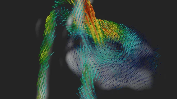
Researchers have developed a new 4D imaging technique that could improve the diagnosis of congenital heart disease before the baby is even born.
The team shared its findings in Nature Communications, noting that this method “could offer exciting new opportunities for assessment of the fetal cardiovascular system in both health and disease.”
The technique turns MRI scans into 4D visualizations of the fetal heart, providing details of major vessels, blood flow and more. Ultrasound has been used to collect such data in the past, but it has long been associated with a series of challenges. This new MRI-based method, on the other hand, produces accurate “movies” that can help clinicians study vessels as small as 5 mm wide.
“If congenital heart disease is detected prior to birth, then doctors can prepare appropriate care immediately at birth, which can sometimes be life-saving,” lead author Thomas A. Roberts, King’s College London, said in a prepared statement. “It also gives parents advance time to prepare, when otherwise the congenital heart disease might have been discovered at birth, which can be very stressful. We are trying to advance fetal cardiac MRI as a way of potentially improving outcomes in congenital heart disease, either by being able to offer a better diagnosis or being able to look for congenital heart disease at an earlier time during pregnancy.”
“The technical challenges that have been overcome by the team in this work represent a massive leap forward in the field of fetal cardiac MRI,” added co-author Kuberan Pushparajah, MD, also of King’s College London. “We will now be able to simultaneously study the heart structures and track blood flow through it as it beats using MRI for the very first time.”
The full analysis is available here.

