Is Interactive Virtual Reality Poised to Deliver a ‘Eureka Moment’ for Cardiology?
When TCT.18’s program planners tasked Saurabh Sanon, MD, with delivering a presentation on interactive virtual reality (VR) that lived up to the conference’s “fascinating lectures” descriptor, he knew his biggest task would be doing justice to the technology. He describes his first time using VR as a “eureka moment.”
He recalls, “I was thinking, there’s no way to convey this to the TCT audience short of giving everyone a set of the 3D glasses and having the screen become 3D compatible.”
Sanon worked with the engineers at EchoPixel, the AR/VR company that had invited him to test-drive the technology six months earlier. For his presentation, they developed a workaround that showed the heart “floating in space, like it appears with the VR glasses on”. Sanon used that visual to show the audience how he has been using VR to refine structural heart disease (SHD) procedures in Florida, where he is the director of the structural heart t ranscatheter therapies program at Palm Beach Gardens Medical Center and a clinical associate professor of medicine at Florida Atlantic University’s College of Medicine.
Speaking with Cardiovascular Business after TCT.18, Sanon elaborated on why he’s convinced VR has the potential to improve patient safety, reduce costs and “provide a new dimension of understanding for cardiac pathophysiology.”
What’s your background and your relationship with EchoPixel?
I trained in structural heart disease and advanced cardiac imaging at the Mayo Clinic in Minnesota. Now I’m at a high-volume SHD center in Florida. Because of the spectrum of SHD cases I encounter, EchoPixel thought I would be a good resource to see how the software works in real life. My role is helping them understand how to use the technology. I’m in no way compensated for using the software.
What was it about interactive VR that intrigued you?
A lot of what structuralists do is in 3D, but our technology has been limited. We see 2D images from X-ray or fluoroscopy and then a similar set of images from TEE or CT. Our expertise is to combine those image sets in our brains and extrapolate that into a 3D image. The better a structuralist can get at understanding 3D space, the better he or she can operate in a cath lab or hybrid OR. That has been the challenge with SHD imaging since the beginning.
When I saw the VR technology and how effortlessly it lets you interact with 3D images, I was totally interested in how the technology might benefit SHD patients. The technology sort of dumbs it down so that any person can look at the data and understand in 3D space what they are looking at.
At TCT.18, you demonstrated how you have used VR to plan left atrial appendage closure (LAAC) procedures. How does VR impact the usual use of TEE or CT images?
The TCT audience saw how I use VR to size and virtually implant a Watchman device (Boston Scientific) using CT images. It took approximately 90 seconds to perform both steps on the VR platform. Of course, I still use TEE and CT, which are considered the gold standard for sizing on all of our cases.
We use the TEE or CT images and sift through them to make the measurements. It might take about 10 to 15 minutes with TEE or even longer with CT. With VR, obtaining the measurements took a fraction of the time.
Besides saving a lot of time on the measurements, another advantage of VR is that you can virtually place the devices in the virtual creation of the patient’s heart, so you can see how it might look once you have done the procedure.
So, you upload the patient’s own images to the VR system? What is VR helping you to work out before the procedure?
I can get a sense of what size device would fit the best and where to puncture the interatrial septum, among other things. Using virtual model delivery sheaths helps me select the right shape of sheath. Short of actually doing the procedure, VR is probably the most realistic simulation of the procedure.
You said VR saved you almost a half-hour with the LAAC case you showed at TCT.18. Is that typical?
Anecdotally, I can tell you it makes the procedure more efficient and saves us some steps where you might try a different sheath size or consider a different puncture location. I’m leading a research study at our site where we’re comparing procedure times with and without VR. That research will give us more concrete numbers. The concept is we’ll end up saving time, make the procedure safer for patients and increase operators’ confidence in the equipment.
Do you use VR for procedures other than LAAC?
Currently, whenever we use VR for case planning or execution, it is as an adjunct to the gold standard, nationally accepted imaging technology. All of our clinical decisions are based on accepted standard-of-care guidelines and techniques. We have applied VR workflows to selecting the right TAVR valve size and seeing the best implantation route. For example, with transcaval TAVR, which is probably the most complex type of TAVR, you can use VR to get a sense of whether the complex transfer between vessels would be safe. I’ve had cases where the VR showed it may be safe for the patient and another where I could see it would be an unsafe proposition. VR helps you get a natural understanding of complex questions.
Another example is paravalvular leak closure. With this technology, you can potentially size the leak and see what plug size you might need to close it. You can get an idea where you might need to enter the heart or where to puncture the septum to get into a certain leak location.
I don’t perform pediatric procedures, but I can see it being useful for congenital heart disease. Anytime you have complex cardiac anatomy or anatomic variability that you need to visualize in 3D or where different devices might have different effects, it’s a good simulation tool.
Would it work in nonstructural settings, such as peripheral artery disease?
Yes, anytime you are dealing with structural formations in the cardiac or vascular beds, it would be useful. It may have applications in noncardiac cases as well.
Could VR be used for patient selection? Might it help you decide not to do a procedure?
Absolutely. I’ve seen that already, where we turned a patient down for a specific therapy because what we saw on the VR was not favorable. A good example might be a patient who had a leak around the mitral prosthesis. He was referred to us for percutaneous closure. Using VR, we found the mitral ring had dehisced from the annulus. Putting in a plug could have worsened the situation. That’s the kind of 3D spatial orientation we can get from using VR that is hard to get with 2D imaging.
Is it difficult to learn how to use this system? Do you need to have a facility for computers?
I may not be the the best person to answer that because I’m a 3D thinker with an aptitude and interest in technology. I used it for a half-hour and knew exactly what to do with it. But the concept is that anyone should be able to figure it out. It’s not like a CT scan, where a radiologist needs months of training to be able to interpret a cardiac CT.
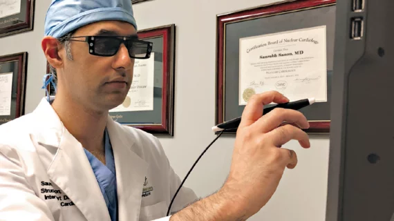
With this interactive VR system, you see an actual heart in front of you and you can use a stylus to spin the heart, move it, interact with it, just like having a physical model. The goal is to make it agnostic of the skill-set of the person using it.
Are there limitations still to be worked out?
I’m still trying to work out the workflows for different procedures. Eventually, as we formalize workflows, we could potentially hit a button and the computer could make measurements automatically. Right now, it’s user assisted. I have to select a certain part of the cardiac anatomy and make the measurement. Then the computer knows how to go to the next step. Further automation is coming.
What about cost and availability?
The structural heart package I’m piloting isn’t commercially available yet; however, the entry-level product for general surgical planning supporting CT, MRI and echo is 510-cleared and costs around $100,000.
At TCT, you predicted VR will become “a ubiquitous part of our workflow.” What timeline do you predict?
Seeing how rapidly the technology is evolving, I would not be surprised if this were very commonly available in the next two to three years.

