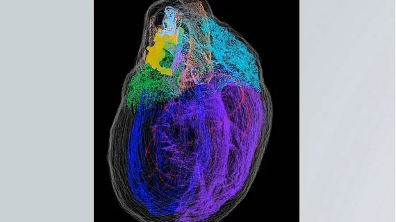
Researchers have developed a detailed 3D map of the intracardiac nervous system (ICN), sharing their findings in iScience. It is believed to be the first map of its kind.
“Autonomic control of cardiac function arises from several integrative centers in the central nervous system, but the final level of neural integration controlling cardiac function lies in the ICN,” wrote lead author Sirisha Achanta, MS, of the Daniel Baugh Institute for Functional Genomics and Computational Biology (DBI) at Thomas Jefferson University in Philadelphia, and colleagues. “Although the heart is known to possess a significant population of neurons, these have not previously been mapped precisely as to their number or extent/position/distribution while maintaining the histological context of the whole heart. This 3D neuroanatomical information is necessary to understand the connectivity of the neurons of the ICN and to develop their functional circuit organization.”
The team turned to knife-edge scanning microscopy and custom-designed software to create and annotate the map of a rat’s ICN, noting that it has provided valuable new insight about neurons inside the heart. Funding for the study came from the National Institutes of Health’s Stimulating Peripheral Activity to Relieve Conditions, or SPARC, research program.
“The ICN represents a big void in our understanding that falls between neurology and cardiology,” James Schwaber, PhD, director of the DBI and a co-senior author of the study, said in a statement. “Our goal was to bridge that gap by providing an anatomical framework of the ICN and a foundation to understand its role in heart health.”
“The only other organ for which such a detailed high-resolution 3D map exists is the brain,” added Raj Vadigepalli, PhD, professor of pathology, cell biology and anatomy at Thomas Jefferson University and a co-senior author of the study. “In effect, what we have created is the first comprehensive roadmap of the heart’s nervous system that can be referenced by other researchers for a range of questions about the function, physiology, and connectivity of different neurons in the ICN.”
What’s next?
Comparisons were also made between male and female rat hearts, and the authors observed sex-specific differences in how the neurons were organized. These subtle differences could potentially help researchers learn more about heart difference in men and women, and the team is planning to craft a similar map of a pig’s heart to explore this potential further.
The team’s ultimate goal, of course, is to use these techniques to produce a similar 3D map of the human heart.
“We’ve created the foundation for an endless possibility of future studies,” Schwaber said.
The full study is available here.

