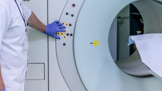
A study published in the latest edition of JACC: Cardiovascular Imaging suggests cardiac magnetic resonance (CMR) imaging can be as effective as measurements of fractional flow reserve (FFR) in evaluating nonculprit lesions after ST-segment elevation MI (STEMI).
The study, penned by Henk Everaars, MD, and colleagues at Amsterdam University Medical Centers, tracked 77 patients with STEMI and at least one intermediate nonculprit lesion. Everaars and co-authors said nonculprit lesions are present in around half of STEMI cases, and CMR presents the unique opportunity to assess a patient’s left ventricular function, infarction size and perfusion simultaneously. Compared to other imaging techniques, CMR is also cost-effective, widely accessible, multiparametric and doesn’t expose patients to ionizing radiation.
“In patients with stable coronary artery disease (CAD), invasive measurements of FFR are considered the optimal index for diagnosing hemodynamically obstructive CAD and guiding revascularization,” the authors wrote in JACC. “Although CMR and FFR are reported to have a high concordance in the assessment of patients with stable CAD, the agreement between CMR and FFR in the evaluation of nonculprit lesions after STEMI is still unknown.”
The team studied the agreement between CMR and FFR in its population of 77 patients, all of whom underwent CMR and invasive coronary angiography in conjunction with FFR measurements one month after their primary STEMI intervention. Only nonculprit lesions with a diameter stenosis between 50% and 90% were considered for the study.
Everaars et al.’s imaging protocol included stress and rest perfusion, cine imaging and late gadolinium enhancement. Fully quantitative, semiquantitative and visual analysis of myocardial perfusion were compared against a reference of FFR, with hemodynamically obstructive lesions defined by an FFR of 0.80 or less.
The authors reported that hemodynamically obstructive nonculprit lesions were present in 40% of patients. Visual analysis revealed an area under the curve (AUC) of 0.74, with a sensitivity of 73% and a specificity of 70%. In semiquantitative analysis, the researchers found the relative upslope of the stress signal intensity time curve and the relative upslope-derived myocardial flow reserve had respective AUCs of 0.66 and 0.71.
Stress myocardial blood flow achieved an AUC of 0.76 with a sensitivity of 69% and a specificity of 77% in the study; myocardial flow reserve achieved an AUC of 0.82 with a sensitivity of 82% and a specificity of 71%.
“As the physiological disturbances resulting from the acute ischemic injury resolve over time, FFR decreases, whereas hyperemic myocardial blood flow (MBF) and myocardial flow reserve (MFR) increase,” the authors explained. “The agreement between CMR and FFR observed in the present study therefore does not reflect failure of either technique but rather underscores that FFR represents a different measure of physiology than hyperemic MBF and MFR.”

