Researchers have designed a new cardiac single photon emission computed tomography (SPECT) imaging system that could potentially deliver images much faster than current models. The team presented its findings at the 2022 annual meeting of the Society of Nuclear Medicine and Molecular Imaging (SNMMI).
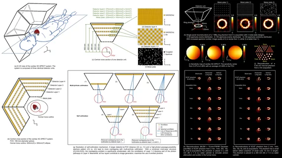
“SPECT is an important noninvasive imaging tool for the diagnosis and risk stratification of patients with coronary heart disease,” co-author Debin Zhang, a doctoral student at Tsinghua University in Beijing, China, said in a statement. “However, conventional SPECT suffers from long scan time and poor image quality as a result of relying on a mechanical collimator. The new SPECT system is capable of performing fast-framed dynamic scans with high quality.”
The team’s proposal revolves around self-collimation, suggesting the restrictions typically associated with a mechanical collimator could be a thing of the past. This self-collimating SPECT nuclear imaging system includes a detector that takes care of both photon detection and collimation, the group explained.
The researchers estimated that this new-look system could produce images 10 or even 100 times faster than the SPECT systems currently being used for cardiac imaging. Its average sensitivity right now is 0.68%, they noted in their presentation.
“The technology proposed in this work may drive a paradigm shift in all single-photon-emission-based molecular imaging and nuclear medicine technologies,” co-author Tianyu Ma, PhD, an associate professor of Engineering Physics at Tsinghua University, said in the same statement. “The new detector design opens up a broad range of possibilities for development of new imaging systems with better image quality, higher speed, and better diagnostic accuracy in molecular imaging.”
The full abstract presented by Zhang et al. is below:
Abstract 250. “Feasibility study of a self-collimating SPECT for fast dynamic cardiac imaging.” Debin Zhang, Xingchun Zheng, Yifan Hu, Zhenlei Lyu, Yaqiang Liu, and Tianyu Ma, Tsinghua University, Beijing, Beijing, China; Zuo-Xiang He, Beijing Tsinghua Changgung Hospital, Tsinghua University, Beijing, Beijing, China; and Rutao Yao, University of Buffalo, State of New York, East Amherst, New York.
More information about SNMMI 2022 is available here. Prior coverage is available here.
Related Cardiac Imaging Content:
7 ways cath labs can work through the contrast media shortage without delays
‘Image of the Year’ highlights the predictive power of a new PET imaging agent

Michael has more than 16 years of experience as a professional writer and editor. He has written at length about cardiology, radiology, artificial intelligence and other key healthcare topics.